Protein and multiomics spatial applications with CFP
All assays are available as manual kits or fully automated on the upcoming S-Light™ instrument
CFP mIF Assay
A.
B.
Panel A: Same slide 10-plex CFP staining performed on FFPE mouse brain section using the Biocare ONCORE Pro X stainer. FFPE BALB/c mouse brain with a view of the hippocampus region. Scale bars =100 µm.
Panel B: Multi-rounds CFP staining of protein markers (12 shown) on the same slide FFPE human lung squamous cell carcinoma. Scale bars =100 μm. This single slide high-plex profile provides more actionable immune context than conventional IHC panels.
RNA ISH Assay
While RNA ISH assays may be more expensive and complex than IHC/IF, its advanced capabilities and detailed insights into gene expression make it a valuable tool for many research and diagnostic applications.
Leading RNA ISH assays are highly sensitive and specific for detecting target RNA molecules, even at low expression levels
RNA ISH allows for studies on targets not suitable for using antibodies such as secreted molecules, non-coding RNA
Detect viral and bacterial nucleic acids in tissue samples
Biomarker validation on animal tissues samples antibody availability is limited
Detection of pathogen and biomarkers on animal tissues.
FFPE HeLa cell pellet stained with Molecular Instruments 2-Plex HCR Pro with MALAT1 and PPIB probes
FFPE human normal breast stained with HCR Pro with PPIB probe
Protein/RNA Co-Detection Applications
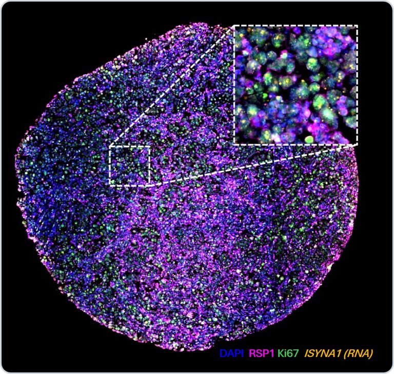
ISYNA1 mRNA were visualized together with RSP1 and Ki67 protein signals, enabling transcript-to-phenotype assessment on the same FFPE section.
CFP RNA and protein co-detection:
CFP enables multiomics detection combining multiplex protein detection and RNA detection from leading RNA ISH providers. The multiomics approach offers researchers flexibility in experiment designs and provides comprehensive multimodal information that helps unveil complex biological processes.
CFP is compatible with 10x Genomics Xenium assays. It enables post-Xenium 10+ plex protein detection using the proprietary cleavable TSA based fluorophores.
*Post-Xenium slide was provided by Biochain
CFP multiomics applications:
Applied downstream of single-cell RNAseq, spatial transcriptomics and other spatial proteomics studies
Use RNA probes to replace unavailable or unspecific antibodies
Perform studies on targets not suitable for using antibodies such as secreted molecules
Offers an orthogonal way for biomarker co-expression and cross-validation in tumor micro-environment (TME), gene therapy and infectious diseases studies
Brightfield Multimodal/Multiomics Applications
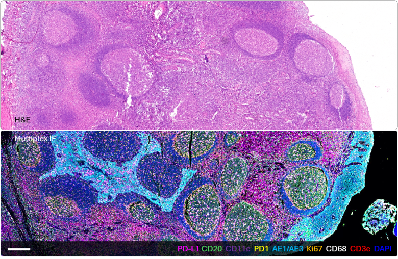
Same slide molecular signal + morphology:
8 protein markers across 2 rounds
Direct IF-to-H&E comparison on the same slide
Reduced uncertainty from serial-section variation
Supports morphology-guided spatial analysis
Co-register multiplex IF with post-IF H&E on the same section. Scale bars =250 μm.
Multimodal readouts, One flexible workflow:
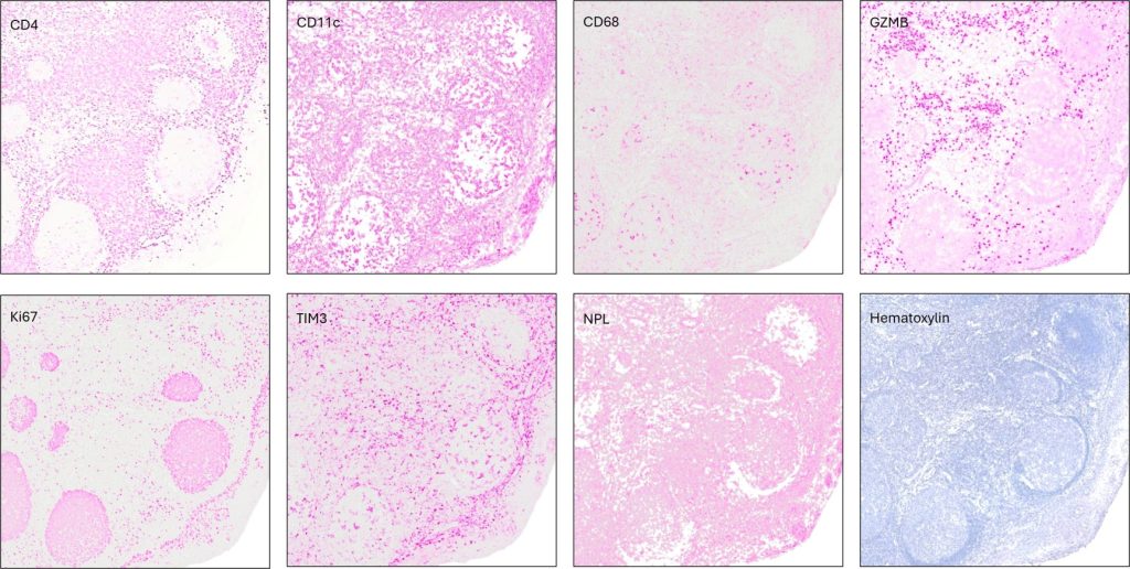
Sequential staining demonstrated repetitive detection of immune and functional protein markers, followed by Vector Laboratories’ Narcissus pseudonarcissus lectin (NPL) for glycan-associated tissue features.
CD4, CD11c, CD68, GZMB, Ki67, and TIM3 were sequentially evaluated. The same tissue section was subsequently stained using Narcissus pseudonarcissus lectin (NPL) from Vector Laboratories to assess glycan-associated tissue features. Hematoxylin staining was also used to visualize tissue morphology.